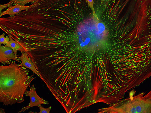
Trabecular Meshwork Cell (detail)  This is the winning image for North America -- IN Cell Image Competition. It shows the internal structure of a single trabecular meshwork cell. In this image, DNA has been stained blue, so the large clumps of blue just above centre are the cell's nucleus. Red lines are filaments of actin spread throughout the cell, while the green patches at their tips are the focal adhesions. In my opinion, the two facts listed below are the most important facts you need to understand about glaucoma. Fact #1: The trabecular meshwork is a contractile tissue. So is the uveoscleral (nonconventional) pathway. Elaboration: The trabecular meshwork is considered the primary site where the fluid (aqueous humor) leaves the eye, and it is a crucial determinant of intraocular pressure. Aqueous humor from inside the eye must flow through the trabecular meshwork to reach Schlemm’s canal where it can flow freely into the aqueous veins. The trabecular meshwork possesses smooth-muscle-like properties. The trabecular meshwork can be induced to contract and relax. When it is contracted, resistance to the fluid flow increases (leading to increased intraocular pressure). The uveoscleral pathway is a pathway directly through the muscle fibers of the base of the ciliary process. The spaces in between those muscle fibers are also closed down when this tissue contracts (again, leading to increased intraocular pressure). Therefore, when there is contraction of these tissues, both points of exit for the aqueous humor are restricted. BTW, ligaments from the muscles of the ciliary process also extend into the trabecular meshwork. Fact #2: All glaucomas have a final common pathway of retinal ganglion cell death involving low-grade inflammation and oxidative stress (as well as mitochondrial dysfunction and glial hyperactivation, both of which have a relationship to inflammation and oxidative stress). In my opinion, these are the two most important facts to understand about glaucoma. Why is that so? Look for a series of articles to continue this discussion on FitEyes.com. (Image source: http://www.newscientist.com/gallery/dn16308-inner-workings-of-cells)
Filed Under (tags):
- dave's blog
- Log in or register to post comments

